Anatomy Of Chest Bone / Bones And Joints Of The Thorax - Bones support and protect the body and its organs.. Pathology of the heart, mediastinum, lungs and pleura. Human chest bone structure parts of the chest bones. Learn about this topic at kenhub! Inserts/attaches on the humerus/upper arm. Identify the following structures on the lateral chest radiograph:
Identify the following structures on the lateral chest radiograph: These joints fuse together in adulthood. Anatomy of the chest wall. Pathology of the heart, mediastinum, lungs and pleura. Bone basics and bone anatomy.
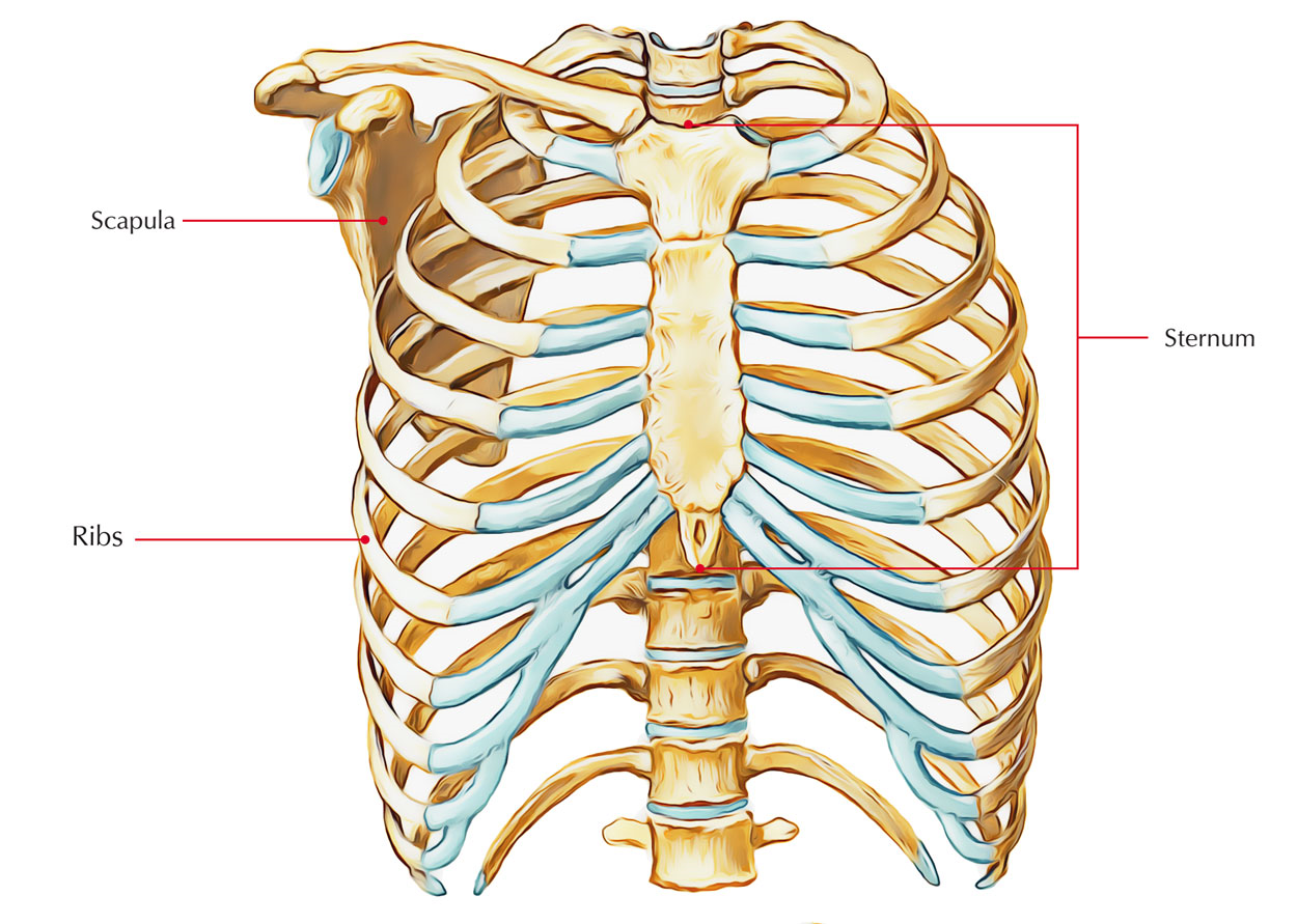
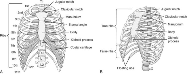
Rib Cage Anatomy Function Britannica from cdn.britannica.com They are always longer than they are wide the vertebrae are irregular bones. The manubrium, sternal body, and xiphoid process. It can help you understand our world more detailed and specific. Bone of chest and their parts. This webpage presents the anatomical structures found on wrist mri. Your rib cage, for example, acts like a shield around your chest to protect important organs inside such as your lungs and heart. The chest can be split into two parts; These joints fuse together in adulthood.
Despite this it is easy to overlook important abnormalities of the bones which may be very subtle.
The ribs meet at an acute angle at the sternum, the costal cartilages thicken like beads at points of their transition to bones (rachitic beads). Where is the sternum found. Bone comprises the structure of the skeletal system and provides lever arms for locomotion. The pec major attaches on the humerus. And we want to know some borders about it. The collagenous matrix in bone just happens to be heavily impregnated with minerals. Anatomists talk about both bone and bones. A collection of anatomy notes covering the key anatomy concepts that medical students need to learn. Upper segment of sternum, flattened roughly triangular bone, o… the bony structure that forms the middle portion of the sternu… Identify the following structures on the lateral chest radiograph: Read the article where all aspects of bone anatomy and physiology are dicussed in detail. The manubrium, sternal body, and xiphoid process. The former is a type of connective tissue made up of cells suspended in a matrix:
Inserts/attaches on the humerus/upper arm. Despite this it is easy to overlook important abnormalities of the bones which may be very subtle. And we want to know some borders about it. We hope you will use this picture in the study and helping chest and abdominal cavities with some organs removed. The pectoralis major and minor.
Easy Notes On Sternum Learn In Just 4 Minutes Earth S Lab from www.earthslab.com Where is the sternum found. All the bones in the body can be described as long bones or bone tissue. Chest bone, ribs, lung, heart, xiphoid process. The former is a type of connective tissue made up of cells suspended in a matrix: Flat bones form by membranous bone formation, whereas long bones are formed by a combination of endochondral and membranous bone formation. It can help you understand our world more detailed and specific. It describes the theatre of events. Sesamoid bones are generally small, flat and have an apex at one end.
Inserts/attaches on the humerus/upper arm.
It describes the theatre of events. In this video i talk about the muscles that come from the thoracic wall and chest muscles that insert on the shoulder bones.✅. Reference database of normal imaging from birth to age 16. Upper segment of sternum, flattened roughly triangular bone, o… the bony structure that forms the middle portion of the sternu… Human chest bone structure parts of the chest bones. They are always longer than they are wide the vertebrae are irregular bones. An overview of the anatomy of the hand, including the bones of the hand, muscles, blood supply and nerve supply. Bone basics and bone anatomy. Long bones are categorised by their tubular shaft (diaphysis) with a rounded end (epiphysis) on each end. The pectoralis major and minor. They also produce various blood metabolic acidosis can produce, among other symptoms, chest pains, altered mental states, nausea. The reason why i do this relates back to the anatomy of the pec major. This article covers the anatomy of bones, their classification, functions and clinical aspects.
Language and terminology for the study of the anatomy of the thorax. Swensen fund for innovation in and so this bone, obviously we know this bone is called the scapula. Bone of chest and their parts. It describes the theatre of events. Your rib cage, for example, acts like a shield around your chest to protect important organs inside such as your lungs and heart.
Bony Thorax Chest And Abdomen Radiology Key from radiologykey.com The wrist consists of multiple joints where the bones of the arm and hand meet. Labeled chest radiographs teaching radiologic anatomy with a level of detail appropriate for medical students. An overview of the anatomy of the hand, including the bones of the hand, muscles, blood supply and nerve supply. Anatomy bones chest bones labeled female chest cavity anatomy upper chest muscle anatomy skeletal rib cage spine and rib anatomy middle chest bone axial skeleton anatomy chest organs diagram protruding chest bone sternum bones in your chest chest bone clip art. Learn about this topic at kenhub! This webpage presents the anatomical structures found on wrist mri. 12 photos of the anatomy bones chest. Long bones are categorised by their tubular shaft (diaphysis) with a rounded end (epiphysis) on each end.
Labeled chest radiographs teaching radiologic anatomy with a level of detail appropriate for medical students.
Bone comprises the structure of the skeletal system and provides lever arms for locomotion. Atlas of anatomy of the human body: When a patient flexes the neck forward, the prominent process is usually that of the 7th cervical. All of the anatomical and important histological facts about the bones, together with the clinical relations, are going to be desrcibed in this article. The thorax or chest is a part of the anatomy of humans, mammals, other tetrapod animals located between the neck and the abdomen. A collection of anatomy notes covering the key anatomy concepts that medical students need to learn. Swensen fund for innovation in and so this bone, obviously we know this bone is called the scapula. This anatomical midline can be useful in assessing for symmetry in breast augmentation or in performing a median sternotomy. Right upper anatomy is to physiology as geography is to history: 12 photos of the anatomy bones chest. The chest can be split into two parts; Bone of chest and their parts. Inserts/attaches on the humerus/upper arm.
The chest can be split into two parts; anatomy of chest. Flat bones form by membranous bone formation, whereas long bones are formed by a combination of endochondral and membranous bone formation.


0 Komentar